AURION Ultra Small Immuno Gold Reagents and AURION R-Gent SE-EM
Application Example 1
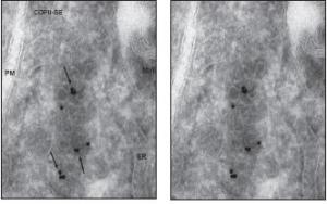
ER exit site in 60 nm-thin cryosection of Hepg2 cells, labeled for COPII (primary antibody against sec23 was obtained by ABR) and detected with Fab-goat-anti-rabbit, conjugated to ultra-small gold, silver enhanced for 30 minutes (from Aurion).
The arrows point to labeled COPII-coats on vesicular and tubular membranes, which are located close to the ER.
The information of a thin section is not sufficient to conclude how the membranes are related to each other- if they are still connected to the ER, or if they are free.
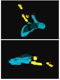
Therefore we performed 3D electron tomography on 400nm thick cryosections, which were labeled similar for COPII. (see next picture).
PM= plasma membrane
MVB= multi-vesicular body
Bar = 100 mikrometer
2 views of a model of a COPII-labeled ER-exit site, resolved from 400nm thick cryo-sections of Hepg2 cells, labeled like described for the ultrathin section before.
Note that the labeling for COPII is assessable throughout the section.
ER=light blue
Free membrane carriers of vesicular and tubular shape, partially labeled for COPII=yellow
COPII=silver enhanced-red
Courtesy of: Dagmar Zeuschner, Judith Klumperman (Department of Cell Biology, UMC Utrecht, The Netherlands) and Willie Geerts, Abraham Koster (Molecular Cell Biology, Utrecht University, The Netherlands).
AURION Ultra Small Immuno Gold Reagents and AURION R-Gent Silver Enhancement
Application Example 2
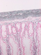 ImmunoGold Silver Staining of laminin in paraffin embedded rat intestine using rabbit polyclonal anti laminin antibody and GAR IgG Ultra Small. Silver enhancement with Aurion R-Gent silver enhancement for 25 minutes. Section is counterstained with nuclear fast red.
Courtesy of Ronnie Wismans, Radboud University Nijmegen Medical Centre, Nijmegen, The Netherlands
AURION Conventional Reagents
Application Example 1
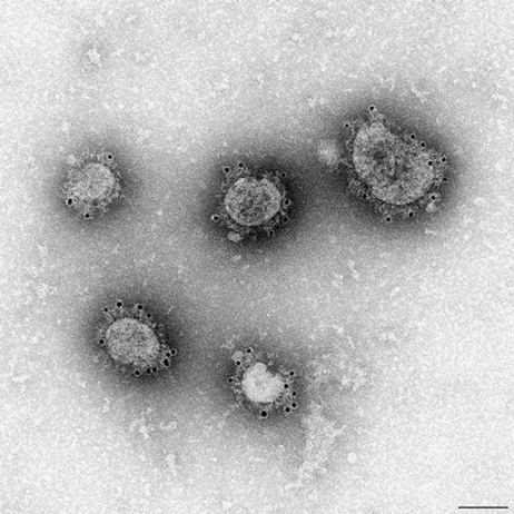
Immunogold labeling of Middle East Respiratory Syndrome-coronavirus (MERS-CoV).
MERS virus is grown in VERO cells via a grid cell culture technique. Primary antibody is a camel anti MERS serum which reacts with the glycoprotein spikes of the virus. Detection with Protein A 10nm. After incubation with the gold reagent specimens are fixed in glutaraldehyde and negative stained with Nano-W.
Courtesy of Sandra Crameri, Australian Animal Health laboratory CSIRO, Geelong, Australia
AURION Conventional Reagents
Application Example 2
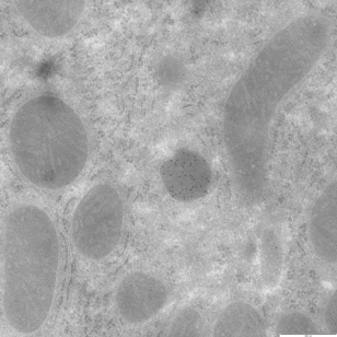
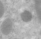 Immunogold labeling of peroxisomes in Tokuyasu sections of perfusion fixed rat liver tissue using 2% paraformaldehyde + 0.1% glutaraldehyde. Catalase was detected using a 1/500 dilution of rabbit anti catalase (Rockland) and Goat anti Rabbit 10nm gold conjugate diluted 1/20.
Specimens courtesy of Torben Ruhwedel, Max-Planck-Institute of Experimental Medicine, Neurogenetics EM, Göttingen, Germany
